The History of Biomedical Imaging
Sep 23, 2020

Biomedical imaging dates to the late 1800s from the discovery of X-rays. The founder, Wilhelm Rontgen, a physicist introduced the idea of radiographs, an image composed from radiation, emitted onto a screen. The 1800s was an era of modern medical imaging, in which real-life images are created from the internal body excluding dissection. The expected output from image scanning ranged between 5 and 11 minutes and can degrade a patient 50 times more radiation compared to today’s X-ray process, which can take milliseconds to process. Over time, the rise of recording media strengthened the quality of X-rays. This revolutionary movement sparked a “growing interest in radiology” amid the Second World War. In the year 1946, fluorescent screens alongside special glasses were being implemented for doctors to detect X-ray images in real life. This gave doctors a glimpse of each integrated X-ray beam, for conceiving unwanted radiation exposure. George Sc Hoenander integrated the first film cassette changer that granted a series of cassettes disclosed for a movie frame rate of 1.5 cassettes per second. In 1953, this procedure had been upgraded to allocate up to 6 frames per second by employing a peculiar cut film changer. Afterwards, in 1955, the X-ray image intensifier was flourished for choosing an exposure of X-ray scanning using a camera and monitor alongside nuclear medicine studies. Moreover, nuclear medicine, also called radionuclide scanning, required low-level radioactive chemicals entering the body. During the 1960s the establishment of ultrasound scanning, generated images under the skin of a human body, without causing damage to the internal organs, and was primarily targeted for pregnant women. “The development of sonar during World War II allowed this to become a reality.” In the early 1970s, a huge leap in digital imaging, led to the creation of the CT Scanner, which had become extensively accessible. Computed tomography scanners, also known as CT Scanner, changed the digital imaging process by introducing major advantages over standard X-ray scanning. CT Scanner has the competence of enlarging images using technology, lower X-ray doses, serenity for storing records and the capability of using computers for evaluating images and recognizing tissues in the human body. Before the move to a computed tomography scanner, these visualizations were either direct, via surgery, or indirect, exacting mental rehabilitation. With the introduction of new three-dimensional (3D) and four-dimensional(4D) medical imaging modalities, this counteracted the need of natural dissection or hypothetical association of anatomy and administer compelling new opportunities for medical examination along with biological analysis. This visualization phenomenon in medicine can extend beyond just singleton molecules and cells, and through an assortment of tissue interfaces alongside integrated organ systems and selected body parts. When the new imaging techniques came into effect in the late 1970s, doctors concluded that they desired ways to analyze images for enlarging the advancement of new imaging techniques. This led to multiple analyses of new methods in medical image analysis. During the 1980s, adopters of magnetic resonance imaging (MRI) attracted scientists and engineers for its images that acknowledge multiple anatomies. This process was an expensive venture. Nevertheless, at the same time, ultrasound was the standard use for producing top-consummation scanners, meanwhile, vendors admitted that clients are selfish for collecting the indispensable money geared towards research purposes. The goal of this research is to provide a clear explanation on the history of biomedical imaging and illustrate different trends of computational applications used in medical diagnosis. Entering the twenty-first century, the science and technology behind biomedical imaging is progressing apart from the standard morphologic and metabolic precedent. As a result, this transition is prospered to adjust patient management and correspond with clinical conclusions. Similarly, as our molecular understanding of the diseases expands, biomedical imaging must also derive onto the molecular level in order to administer a custom-made approach for each individual clinical probe scenario.
Current Situation
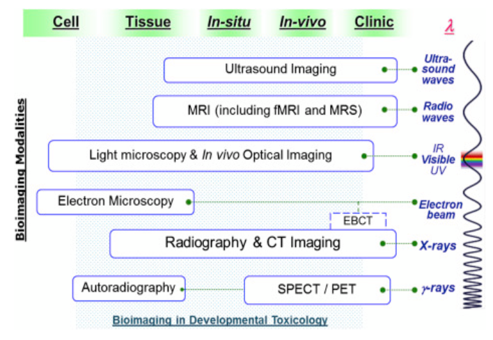
Considering the start of Victorian Times, the exploration of biomedical imaging is geared towards finding noninvasive ways to glance into the human body by accepting light emitted photons. With the start of a new decade, new innovations introduced in the market are conating to produce healthcare more adequately. Nevertheless, the healthcare industry is conating to make healthcare further compelling. The top four compelling technology innovations prone to shape medical diagnosis include, “Faster acquisition enables improved image quality. Gain in optimal dose efficiency. 2D image capture evolving to 3D. AI as a supplemental lens for medical image analysis.” To fully understand the utilization behind biomedical imaging, a microscopic visualization of cells, and molecules must be acquainted by. Scientists must be willing to undergo training for each individual machine, both in terms of hardware and software knowledge. Current generation X-rays require an insight in solid-state electronics, whereas, previous propagation included each result to be recorded on film cassettes. Biomedical technologies have presented multiple innovative approaches towards multidimensional detection, cell imaging visualization, and developmental toxicology. Below is an example of the current advancement in development toxicology.
In addition, there are other proprietary biomedical imaging techniques, including “optical imaging(OI), magnetic resonance imaging(MRI), positron emission computed tomography(CT), and conventional radiography applied clinically.” It is still unclear that researchers have yet found the ideal result. Below is a graph showcasing the number of computer tomography scanners in selected countries as of 2017, powered by Statista.
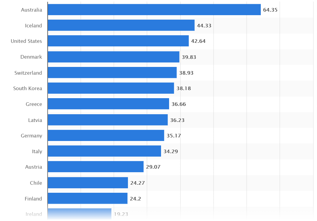
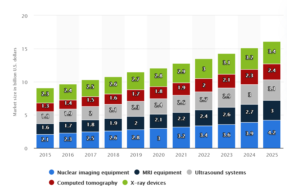
The numbers displayed to the right of each country are expressed per million population. On the contrary, the amount of medical imaging devices in the United States is expected to increase in market size. Due to a shift in testimony, researchers have begun to experiment tests by applying label-free chemical contrast signalling special chemical interactions. Furthermore, a 3D dimensional multiphoton vibrational imaging technique pampers the microbiology of each tissue in order to analyze the side effects of ingesting drug delivery from label-free chemical contrast. Even if the research does not successfully sell to the market, multiple medical facilities will tailor their funds towards a more tailored equipment. Without a doubt, robotic companies are pouring assets into possible funding towards biomedical research that involve the assistance of fully autonomous imaging equipment. The global medical robotics market is rapidly expanding at a rate of 18.2% annually, while maintaining an ambition to support patients. Robotic companies are striving to endeavour control, and flexibility in assisting a patient's scan, medical diagnosis or any available accommodation all while reducing common symptoms including pain, reduced blood loss, and transmission plus the help of machine learning techniques. Switching gears, the total U.S. market cap is about 9.7 billion U.S. dollars, 2.4 billion of which were achieved from X-ray devices. Below is a graph representation of medical imaging market size in the U.S. from the year 2015, up to 2025, by product.
History
The starting point of biomedical imaging dates back to the year 1895 with the revelation of X-rays. X-ray imaging is done through a transmission-based technique, to demonstrate that a connection over a patient is distinguished “either by film or an ionization chamber on the opposite site of the body.” After one year, X-rays become an oddity, and doctors across the globe start adopting the technology. By the year 1955, a Scottish physician examined the adoption of ultrasound for medical diagnosis. Immediately after, the first fiber-optic semi-flexible gastroscope is acquired. In 1958, a paper on medical diagnostic ultrasound became publicly available. Following the research on ultrasound, the endoscope is audited in the stomach of a patient. At the start of 1957, the CT Scanner was introduced in the field of science and technology. Computed Tomography(CT) Scanner is the action of scanning a body by applying the basis of an X-ray. As a result of this, in 1971, the first ever CT scan was implemented on a patient’s brain. Eventually in 1973, an American Chemist with the help of a nuclear magnetic resonance data and computer tomography arithmetics, created the first magnetic resonance imaging(MRI) image. Then in 1974, the creation of the positron emission tomography(PET) camera was introduced alongside the inaugural whole-body system geared towards human and animal inquiries. Shortly after, the first ever MRI body scan is completed on a hominid employing an MRI computer. Ultrasound scanning rises in popularity following the adoption of MRI scanners inside hospitals during the early 1980s. Time magazine grants PET/CT scanners as the medical innovation of the year 2000. Lastly, in September of 2014, the university of Canterbury was prone to $12 million for developing the world’s first colored X-ray scanner.
What's Next
Concisely, Richard A.Robb dedicated his time in researching the theory behind three dimensional biomedical imaging. His analysis towards the medical exertion, besides single photon emissions, stated the benefits of discrete spect systems, rotating gamma camera systems, and clinical applications.These studies briefly explained the primary ambition of image reconstruction processes. Furthermore, Andrew Webb discusses various strategies of biomedical imaging, which correspond to nuclear medicine, X-ray imaging and computed tomography, ultrasonic imaging, magnetic resonance imaging, and general image characteristics. He describes each topic precisely by theory and further explanations with vivid illustrations. In other words, his description on computed tomography emphasizes the amount of radiation dose a patient might experience when performing image processing. On the contrary, an article written by Greg Freiherr provides a prototype of the first computed tomography scanner. The University of Waikato in New Zealand, demonstrates a timeline of each decisive action of biomedical imaging from the Victorian Times up to the new decade. This timeline presents major developments in biomedical imaging. Meanwhile, Image Science expresses their interests on time-lapse imaging by concluding a legitimate understanding of the cellular and molecular processes, which undergo living organisms. Experiments that involve time-lapse imaging can produce outcomes that are up to five-dimensional(5D) image data sets. Moreover, Science Direct has uploaded a series of pdf files explaining the structures of achieving, transforming, and envisioning functional images of living objects or systems. The study has been posted in 2018, by multiple contributions from several authors interested in the study of biomedical imaging. Last, but not least, IEEE Engineering in Medicine & Biology Society portrays the science and technology trailing each conceived sensor, instrumentation and software implementation from X-rays and computer tomography scanners.
Conclusion
In conclusion, biomedical imaging can be a compelling system for visualizing the internal structure of the body, and can reduce the proneness of diseases. With the adoption of new technology, today’s imaging processes feature extraordinary aspects of biological processes. Amidst X-ray innovations, CT Scanners are widely adopted for precluding screening on diseases. Performance with ultrasound imaging is abstinence, compact, and reasonable for providing design and behavior in a programmable approach. The discussion of biomedical imaging can proceed with the expansion of different medical equipment and their impacts on image scanning. What are some of the drastic differences in machine operation and how can one benefit from the other when it comes to medical processing capabilities? In order to fully comprehend an organism from differentiating between a healthy and sick body, we must take into consideration metabolic figures, both big and miniscule in sync with their communication lineation. Without a doubt, label-free approaches can be taken in deliberation by adopting plasmonic microscopy.